
Thermography, also known as Digital Infrared Thermal Imaging (DITI), is a noninvasive, no contact or radiation, clinical imaging procedure showing the thermal abnormalities present in the bodies of children, men and women:
- To help in determining the cause of pain
- To aid in the early detection of disease and pathology
- To evaluate sensory-nerve irritation or significant soft tissue injury
- To define a previously diagnosed injury or condition
- To identify an abnormal area for further diagnostic testing
- To follow progress of healing and rehabilitation
DITI has been recognized as a viable diagnostic tool since 1987 by the AMA Council on Scientific Affairs, the ACA Council on Diagnostic Imaging, the Congress of Neuro-Surgeons in 1988 and in 1990 by the American Academy of Physical Medicine and Rehabilitation. DITI was FDA approved in 1982 and aids in the diagnosis and prognosis, as well as monitoring therapy progress, for conditions and injuries, including:
- Arthritis
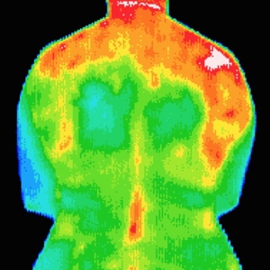
- Back Injuries
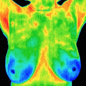
- Breast Disease
- Cardiac Dysfunction
- Carpal Tunnel Syndrome
- Dental and TMJ
- Diabetes
- Digestive Disorders
- Disc Disease
- DVT
- Epicondylitis
- Fibromyalgia
- Headaches
- Inflammatory Pain
- Limb Ischemia
- Muscle Injuries
- Nerve Damage
- Raynaud’s Disease
- RSD (CRPS)
- Skin Cancer
- Sprain/Strain
- Stroke Screening
- Synovitis of Joints
- Tendon Sheaths
- Unexplained Pain
- Vascular Disease
- Whiplash
And many more conditions! Take a look at the examples below:
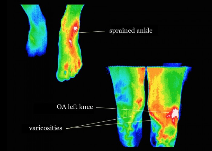 Varicosities

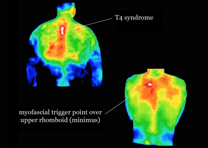

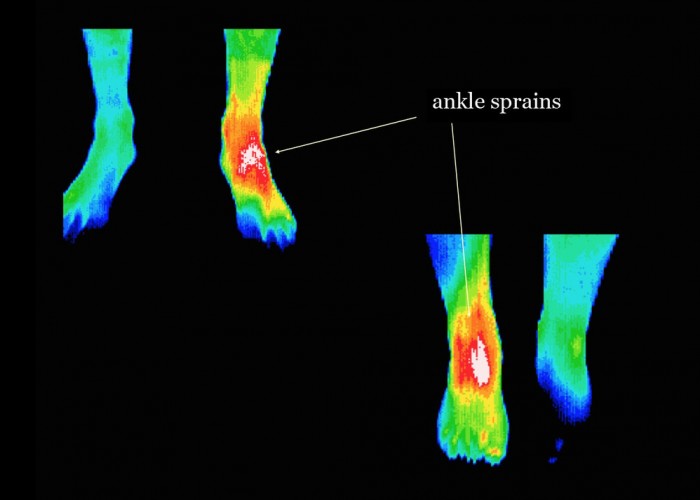
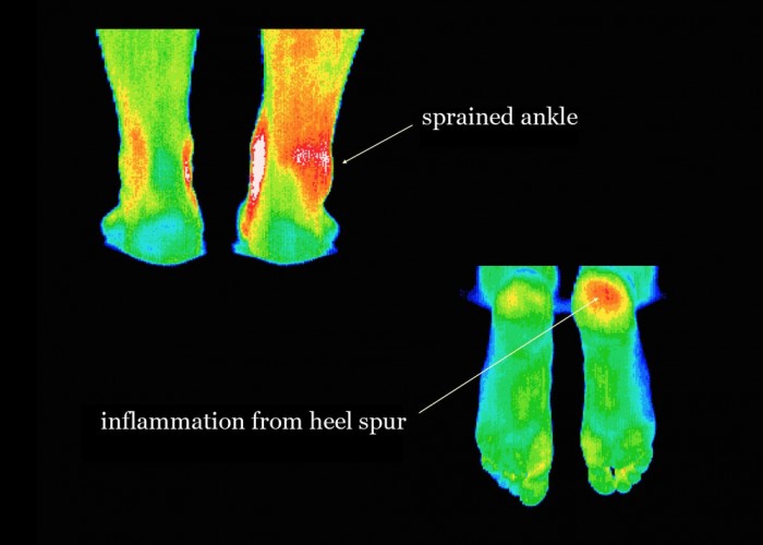
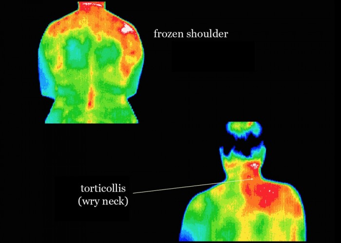

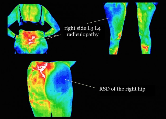
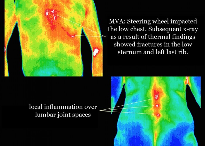
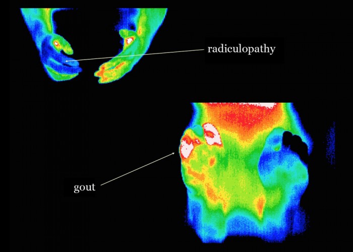
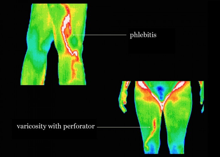


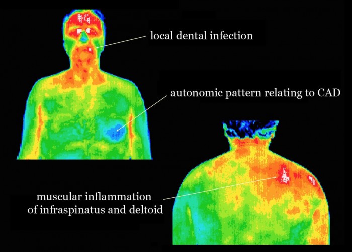
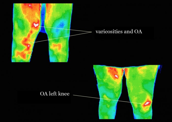


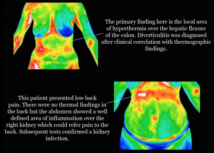


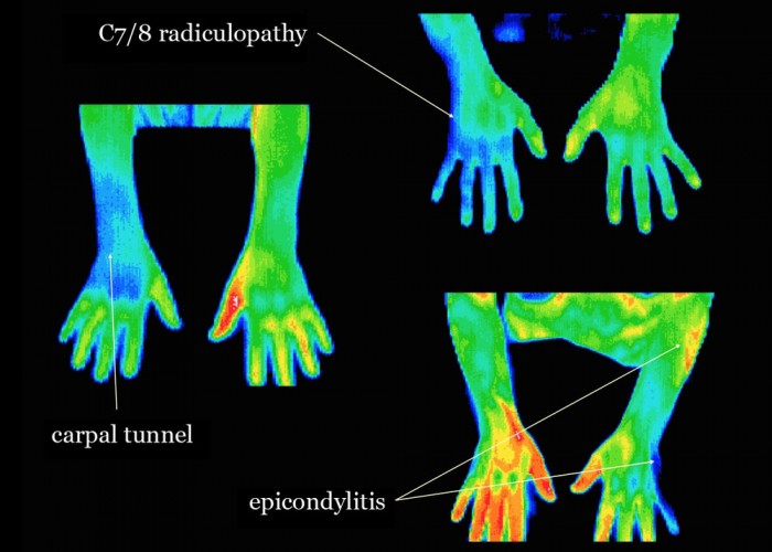
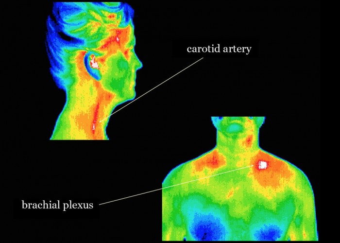


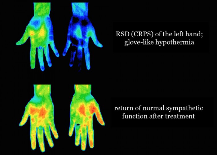


Medical DITI can graphically display the very subjunctive feeling of pain by objectively displaying the changes in skin surface temperature that accompany pain states.
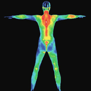
DITI is a noninvasive diagnostic technique that allows the examiner to visualize and quantify changes in skin surface temperature. An infrared scanning device is used to convert infrared radiation emitted from the skin surface into electrical impulses that are visualized in color on a monitor. These visual images graphically maps the body’s temperature and is referred to as a thermogram. The spectrum of colors indicates an increase or decrease in the amount of infrared radiation being emitted from the body’s surface. Since there is a high degree of thermal symmetry in the normal body, subtle abnormal temperature asymmetries can be easily identified.
There are many tests of anatomy, such as x-ray, CT, ultrasound and MRI. Thermography is unique in its capability to show physiological change and metabolic processes. It has also proven to be a useful complementary procedure to other diagnostic modalities. Thermography is certainly an adjunct to the appropriate usage of mammography and not a competitor. In fact, thermography has the ability to identify patients at the highest risk and actually increase the effective usage of mammography imaging procedures.
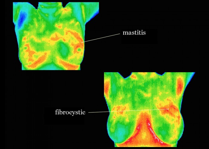
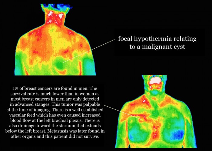
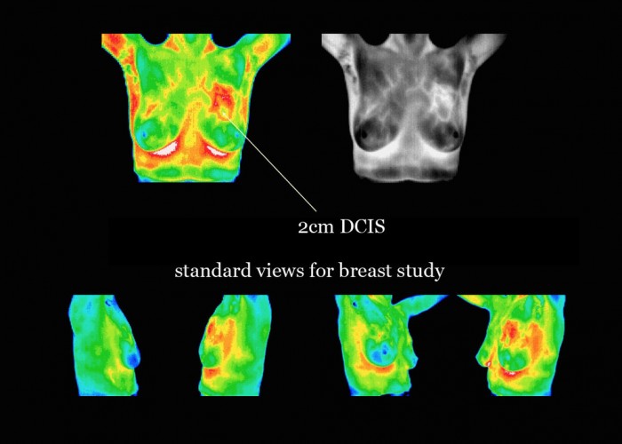
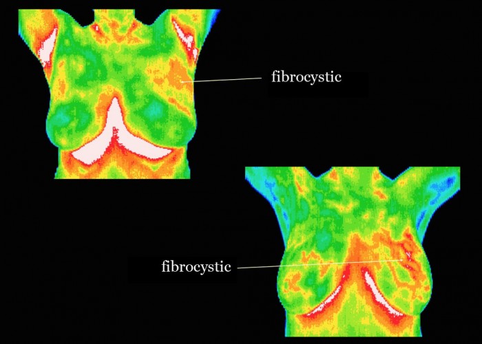
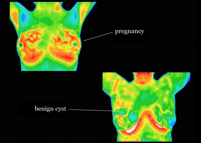
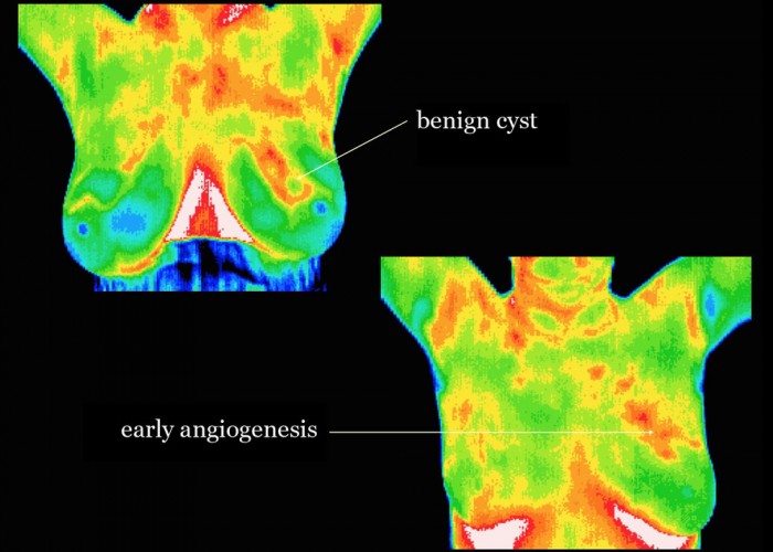
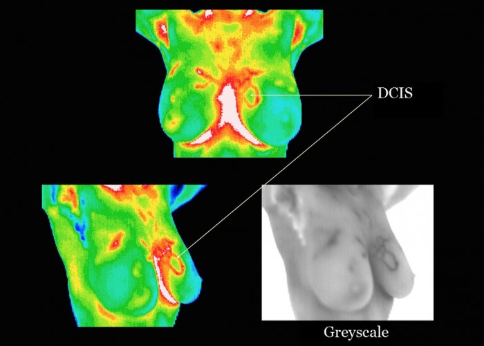
By performing thermography years before conventional mammography, a selected patient population at risk can be monitored more carefully. Then by accurately utilize mammography or ultrasound as soon as possible to detect the actual lesion (once it has grown large and dense enough to be seen on mammography film) can increase treatment options and ultimately improve the outcome.Thermography offers the opportunity to detect breast disease, without radiation or contact, 5-8 years earlier than has been possible through breast self examination, doctor examination or mammography alone. DITI detects the subtle physiologic changes that accompany breast pathology whether it is cancer, fibrocystic disease, an infection or a vascular disease. It is one of the only medical procedures to detect inflammation breast cancer. Thermography, as a physiological test, demonstrates heat patterns that are strongly indicative of breast abnormalities. Once abnormal heat patterns are detected in the breast, follow up procedures including mammography are necessary to rule out or properly diagnose cancer and a host of other breast diseases.
All women can benefit from DITI breast screening. However, it is especially appropriate for younger women (30-50) whose denser breast tissue makes it more difficult for mammography to be effective. Also for women of all ages who, for many reasons, are unable to undergo routine mammography. This scan can provide a ‘clinical marker’ to the doctor or mammographer that a specific area of the breast needs particularly close examination. It takes years for a tumor to grow, thus the earliest possible indication of abnormality is needed to allow for the earliest possible treatment and intervention. DITI’s role in monitoring breast health is to help in early detection and monitoring of abnormal physiology.
Thermograms can be taken of the whole body or just areas being investigated. Our office offers the following thermography scans:
Thermography can offer considerable financial savings by avoiding the need for more uncharted, expensive investigation for many patients.
-Meditherm & ACCN